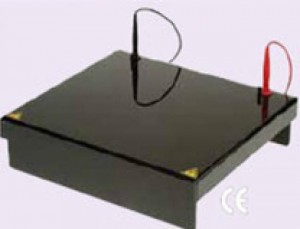
Cleaver Scientific COMET assay tanks are available in four slide formats to study single cell gel electrophoresis (SCGE), a technique made popular by drug toxicology and carcinogenesis studies for the detection and quantitation of DNA damage in cells. Each tank’s robust construction from ebony acrylic ensures that cells remain free of exposure to background light and DNA damage during electrophoresis, while a cooled central platform provides a convenient surface for slide preparation and control of slide temperature during the assay. Following electrophoresis DNA damage may be measured using the highly recommended Comet Assay IV Software from Perceptive Instruments.
F250 Chiller
With PID control ensuring temperature stability within ±0.5°C, the F250 is the perfect recirculating chiller for active slide temperature control. This chiller also benefits from a maximum flow rate of 15 litres per minute and 0.35 bar pressure, while a small footprint and frontal vents allow multiple units to be aligned side-by-side if required. A versatile working temperature range of +5°C to +40°C makes the chiller suitable for use in other applications such as flat-bed isoelectric focusing.
The Comet Assay
Background: First introduced in 1981 to quantify double-stranded DNA breakages in single cells exposed to γ-irradiation, the Comet Assay (or SCGE) has since been adapted to analyse specific DNA lesions and repair processes.
Overview: Following genotoxic insult, such as ionizing radiation, the resultant strand breakage of supercoiled duplex DNA reduces the size of the large genomic DNA from which these strands are separated or drawn out by electrophoresis. The genomic DNA then takes on the appearance of a ‘comet’ as its negatively charged broken ends and fragments migrate towards the anode during electrophoresis.
Method: After exposure to a genotoxic insult cells are suspended within low melting point agarose and embedded within a thin layer of agarose on a microscope slide. Cellular protein is then removed by lysis in detergent, when DNA is allowed to unwind in alkaline conditions before electrophoresis. The DNA is electrophoresed, stained and then analysed using software such as Perceptive Instruments Comet Assay IV.
Results: Microscopy imaging is used to measure DNA fluorescence upon staining. In DNA damaged cells the resultant image resembles a ‘comet’ with the cellular DNA separated into a head and tail. The head is mainly composed of intact genomic DNA, whereas any fragmented or damaged DNA is concentrated within and towards the tail.